Understanding Endometriosis-Induced Pelvic Floor Hypertonia
Deep infiltrating endometriosis (DIE) presents a complex clinical challenge, frequently characterized by chronic pelvic pain, dyspareunia, and notable pelvic floor muscle hypertonia. For physiotherapists treating women’s health conditions, addressing the musculoskeletal components of endometriosis is paramount. Hypertonicity of the pelvic floor muscles often exacerbates pain symptoms, creating a vicious cycle of tension and nociceptive feedback. A recent randomized controlled trial highlights the profound impact that targeted pelvic floor physiotherapy (PFP) can have on these anatomical structures and their associated symptoms.
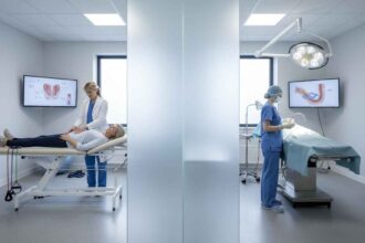
The Role of 3D/4D Transperineal Ultrasound in Assessment
Traditionally, evaluating the efficacy of physiotherapy interventions for pelvic floor dysfunction relied heavily on subjective patient reporting and manual palpation. However, researchers Del Forno et al. (2021) utilized 3D/4D transperineal ultrasound to provide objective, quantifiable measurements of the levator ani hiatus area (LHA). By measuring the LHA at rest, during maximum voluntary contraction, and particularly on maximum Valsalva maneuver, clinicians can precisely observe changes in pelvic floor muscle relaxation and compliance. This advanced imaging technique bridges the gap between functional physical interventions and measurable anatomical outcomes.
Clinical Interventions and Key Study Findings
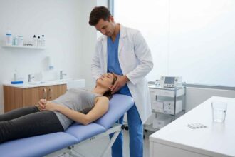
This pivotal randomized controlled trial involved nulliparous women diagnosed with DIE and superficial dyspareunia. The treatment group underwent five individual sessions of pelvic floor physiotherapy, while the control group received no intervention. Four months later, follow-up evaluations yielded compelling clinical results. The primary outcome measure—the percentage change in LHA on maximum Valsalva maneuver—demonstrated a significant functional improvement in the study group (20.0 ± 24.8%) compared to the control group (-0.5 ± 3.3%). This objective increase in hiatal area strongly indicates enhanced pelvic floor muscle relaxation following targeted physiotherapy. Furthermore, subjective symptom reporting mirrored these positive anatomical changes. Women who received PFP experienced a marked reduction in superficial dyspareunia and a statistically significant improvement in chronic pelvic pain.
Implications for Physiotherapy Practice
For evidence-based physiotherapists, this trial underscores the vital necessity of integrating specialized pelvic floor muscle therapy into the multidisciplinary management of deep infiltrating endometriosis. The demonstration that a relatively short intervention—just five targeted sessions—can yield substantial morphological and symptomatic improvements is highly encouraging. It validates the clinical reasoning that alleviating pelvic floor hypertonia directly reduces chronic pelvic pain and pain during intercourse. As practitioners, advocating for objective tracking methods or working in tandem with gynecological sonographers can elevate the overall standard of care, offering tangible proof of treatment efficacy to patients suffering from chronic pain and functional limitations.
References
Del Forno, S., Arena, A., Pellizzone, V., Lenzi, J., Raimondo, D., Cocchi, L., Paradisi, R., Youssef, A., Casadio, P., & Seracchioli, R. (2021). Assessment of levator hiatal area using 3D/4D transperineal ultrasound in women with deep infiltrating endometriosis and superficial dyspareunia treated with pelvic floor muscle physiotherapy: randomized controlled trial. Ultrasound in obstetrics & gynecology : the official journal of the International Society of Ultrasound in Obstetrics and Gynecology. https://pubmed.ncbi.nlm.nih.gov/33428320/