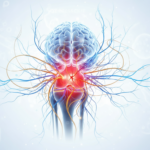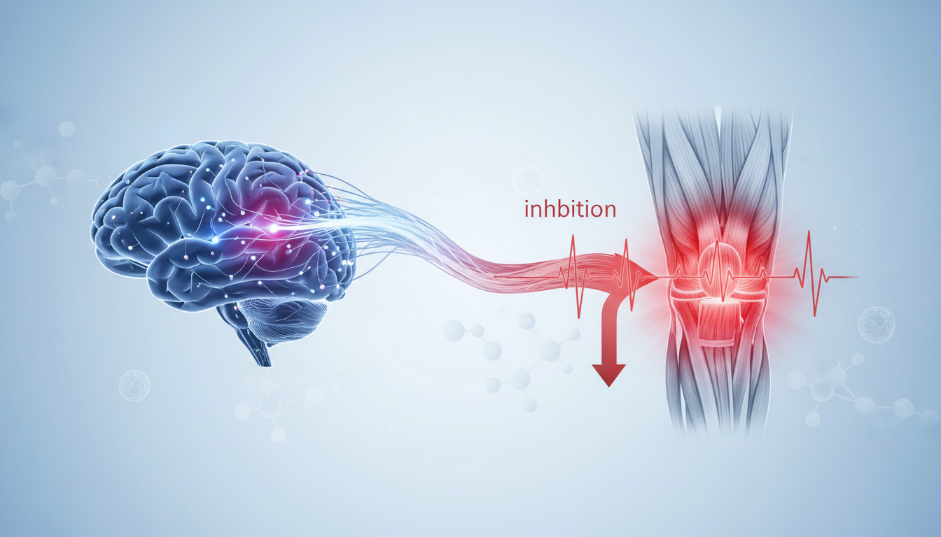
The Neural Component of Patellofemoral Pain
Patellofemoral pain (PFP) is one of the most common musculoskeletal conditions, often presenting as persistent aching in the front of the knee. For decades, rehabilitation focused almost exclusively on local mechanical factors, such as patellar tracking and hip strength. However, emerging evidence suggests that the central nervous system—specifically the corticospinal pathway—may be a hidden driver of the persistent weakness and dysfunction seen in these patients. This neural perspective offers a significant shift in how we understand and treat chronic knee pain.
Study Overview: Mapping the Brain-Knee Link
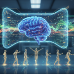
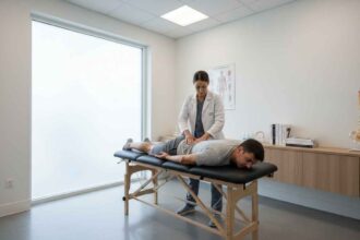
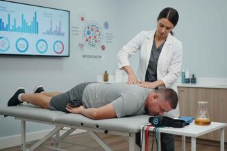
A recent cross-sectional study led by Gevrek Aslan and colleagues, published in the Journal of Sport Rehabilitation, investigated the neurophysiological mechanisms underlying quadriceps impairment. The researchers utilized Transcranial Magnetic Stimulation (TMS), a non-invasive technique that stimulates the motor cortex to measure how effectively the brain communicates with the muscles. The study compared 21 individuals with PFP against 21 pain-free controls, evaluating several key metrics:
- Cortical Silent Period (CSP): A measure of inhibitory activity within the motor cortex.
- Motor Thresholds: Resting and active levels of excitability.
- Quadriceps Strength: Measured via manual muscle testing and specialized biofeedback.
- Self-Reported Outcomes: Including pain intensity, kinesiophobia, and functional capacity.
Clinical Findings: The Role of Inhibition
The study’s most significant finding was that individuals with PFP exhibited a significantly longer cortical silent period (CSP) compared to healthy controls. In neurophysiology, a longer CSP is indicative of increased GABA-ergic inhibition within the primary motor cortex. Essentially, the brain is “braking” the signal sent to the quadriceps. This inhibitory state was found to be bilateral, meaning it affected both the painful limb and the asymptomatic limb in PFP patients. This suggests a systemic neural adaptation rather than just a local response to a specific knee injury.
Impact on Strength and Function
What makes this research particularly impactful for clinicians is the strong correlation between these neural markers and clinical symptoms. The data revealed that a longer CSP was directly associated with:
- Increased Pain Intensity: Higher levels of perceived pain were linked to greater cortical inhibition.
- Reduced Knee Function: Patients with longer silent periods scored lower on functional assessments.
- Lower Quadriceps Strength: Inhibition in the motor cortex was a clear predictor of muscle weakness.
- Longer Pain Duration: Chronic cases showed more pronounced inhibitory signatures.
These findings suggest that the “weakness” observed in PFP might not be entirely due to muscle atrophy or local joint issues, but rather a neural “shutting down” of the muscle’s capacity to activate effectively.
Conclusion: Implications for Rehabilitation
The discovery of impaired corticospinal excitability shifts the paradigm of PFP treatment. If the brain is actively inhibiting the quadriceps, traditional strength training alone may not be enough for all patients. Future rehabilitation strategies may need to incorporate “neuro-priming” techniques, such as neuromuscular electrical stimulation or cognitive-motor training, to reduce this cortical inhibition. By addressing the neural drivers of pain and weakness, clinicians can provide more comprehensive and effective care for those suffering from patellofemoral pain.
APA Citation:
Gevrek Aslan, C., Kilinc, H., Cengiz, B., Turhan, E., & Kinikli, G. I. (2026). Impaired Corticospinal Excitability of the Quadriceps in Individuals With Patellofemoral Pain is Associated With Worse Pain, Function, and Strength. Journal of Sport Rehabilitation, 1-10. https://doi.org/10.1123/jsr.2025-0012
Source: PubMed Central