Ulnar tunnel syndrome, also known as entrapment of the ulnar nerve at the wrist, involves compression of the ulnar nerve as it passes through the ulnar tunnel (or Guyon’s canal) near the wrist. This condition often leads to tingling, numbness, or weakness affecting the little finger and part of the ring finger. When left untreated, it may lead to loss of fine motor control, grip strength, and functional impairment of the hand.
Anatomy and Pathophysiology
The ulnar nerve originates from the brachial plexus and travels down the arm, passing behind the elbow and then entering the wrist through a narrow passageway known as the ulnar tunnel or Guyon’s canal. At this location, the nerve has both sensory and motor branches supplying the ulnar side of the hand, including the little finger and half of the ring finger, as well as intrinsic hand muscles.
Compression of the nerve in this tunnel may arise from a variety of mechanisms: a ganglion cyst or fluid-filled sac pressing on the nerve, fracture or dislocation of the nearby carpal or hamate bones, repetitive pressure on the wrist (as from cycling or tool use), or thickening of surrounding tissues. Over time, the compression may lead to demyelination, impaired nerve conduction, muscle fibre loss, and atrophy of small hand muscles. The combination of sensory and motor dysfunction leads to the typical clinical picture.
Etiology and Risk Factors
Mechanisms
Common causes of ulnar tunnel syndrome include direct pressure on the wrist, especially over the hypothenar eminence; repetitive wrist movements or prolonged wrist flexion/extension; trauma to the wrist bones (for example a hamate fracture or hook of hamate injury); ganglion cyst formation inside the tunnel; and anatomical variations or swelling within the canal that reduce the available space for the nerve.
Risk Factors
- Repetitive wrist or hand use, especially in gripping or pressure-bearing activities (for example, cyclists, manual labourers)
- Previous wrist injury, fracture, or dislocation affecting the carpal or hamate bones
- Ganglion cysts or other masses near the ulnar tunnel
- Wrist anatomy that reduces the space in the tunnel
- Occupations or sports involving prolonged use of tools, handlebars, or devices pressing on the ulnar side of the wrist
- Conditions that predispose to nerve vulnerability (for example, metabolic disorders, inflammation)
Clinical Presentation
Symptoms
Patients typically present with tingling, numbness, or “pins and needles” sensation in the little finger and the ulnar half of the ring finger. These symptoms may occur intermittently at first, often when the wrist is pressed or bent, and may worsen over time to become frequent or constant. Weakness in grip, dropping objects, difficulty with fine movements (such as buttoning, typing,) or a feeling of clumsiness in the ulnar-side hand may be reported. Pain at the ulnar side of the wrist or hand may also accompany the nerve symptoms.
Physical Examination
On examination, there may be:
- Tenderness or sensitivity when palpating the ulnar tunnel region at the wrist
- Sensory impairment in the little finger and part of the ring finger
- Weakness of intrinsic hand muscles supplied by the ulnar nerve (for example, interossei, adductor pollicis) indicated by difficulty spreading fingers or a positive sign, such as Froment’s sign (thumb flexion when pinching paper)
- Muscle wasting of the hypothenar eminence or interossei in more advanced cases
- Tinel’s sign: tapping over the ulnar tunnel may provoke tingling in the distribution of the ulnar nerve
- Observation of hand posture, grip strength, and ulnar deviation or wrist alignment
Because the ulnar nerve can be compressed at multiple points (wrist or elbow), the examiner should also assess for proximal entrapment or other nerve pathologies.
Diagnostic Evaluation
Imaging and Studies
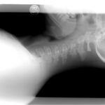
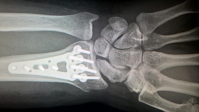
Initial imaging may include plain radiographs of the wrist to identify fractures, bone abnormalities (for instance, of the hamate or carpal bones), and any masses or cysts. Advanced imaging (ultrasound, MRI) can visualise soft-tissue structures, ganglion cysts, or space-occupying lesions within the tunnel and also assess the nerve and muscle changes.
Electrodiagnostic studies (nerve conduction velocity, electromyography) help confirm the diagnosis, quantify the degree of nerve dysfunction, locate the site of compression, and guide treatment decisions by assessing sensory and motor nerve conduction across the ulnar tunnel.
Assessment
A thorough history should clarify onset, aggravating factors (wrist pressure, cycling, tool use), pattern of symptoms, and any previous wrist injuries. Clinical examination and studies allow classification as mild, moderate, or severe nerve involvement, which influences management strategy and prognosis.
Treatment
The goal of treatment is to relieve nerve compression, restore nerve function, preserve hand strength and function, and prevent permanent nerve damage or muscle atrophy.
Conservative Management
For mild or early cases, nonoperative management is the first line and may consist of:
- Activity modification to reduce repetitive wrist pressure on the ulnar side or avoid prolonged wrist flexion/extension
- Use of a wrist brace or splint, especially during sleeping or activities causing symptoms
- Non-steroidal anti-inflammatory drugs (NSAIDs) or other analgesics for symptom relief
- Ergonomic adjustments (such as handlebar padding for cyclists, tool modifications)
- Physical therapy, including nerve gliding exercises, strengthening of hand and forearm muscles, and addressing wrist posture and mechanics
- Monitoring for progression of symptoms, especially muscle weakness or wasting
Surgical Management
Surgery may be indicated in moderate to severe cases, those failing conservative treatment, or when a clear structural lesion is identified (for example, ganglion cyst, fracture fragment). Surgical options include decompression of the ulnar nerve within the tunnel (opening the roof of Guyon’s canal), excision of cyst or lesion, sometimes combined with neurolysis or ulnar nerve transposition to a less vulnerable position. Postoperative rehabilitation is critical to regain strength, range of motion, and function.
Rehabilitation and Follow-Up
After either conservative or surgical treatment, rehabilitation focuses on restoring wrist range of motion, strengthening hand and forearm muscles, improving dexterity, and a gradual return to full activity, including sports or occupational tasks. Periodic follow-up is important to monitor for resolution of sensory symptoms, return of motor function, prevention of recurrence, and identification of any remaining nerve damage or muscle atrophy. In cases of longstanding compression, complete recovery may take months, and residual deficits may persist.
Complications and Prognosis
Potential complications include persistent numbness or tingling, incomplete recovery of grip strength, muscle atrophy of the hand intrinsics, and permanent nerve damage if compression is prolonged. Delay in treatment, severe compression, or associated wrist pathology (fracture, cyst) increases the risk of a poorer outcome. Prognosis is generally favourable when compression is recognised early and managed promptly; full functional recovery is likely in many cases, though mild persistent symptoms may remain.
Prevention and Patient Education
Key preventive strategies include:
- Avoiding prolonged pressure on the ulnar side of the wrist (for example, by using padded gloves or handlebar modifications)
- Minimising repetitive wrist flexion/extension or maintaining the wrist in neutral during sleep or work
- Early evaluation of wrist pain, numbness, or finger tingling, especially among cyclists, tool users, or individuals with previous wrist trauma
- Hand and wrist strength and conditioning, ergonomic assessments of work or sporting equipment, and education on nerve-saving wrist positions
- Prompt referral if symptoms of ulnar nerve compression persist, to avoid irreversible nerve damage
References
Chen, S. H., & Tsai, T. M. (2014). Ulnar tunnel syndrome. The Journal of hand surgery, 39(3), 571-579.
Ginanneschi, F., Mondelli, M., Cioncoloni, D., & Rossi, A. (2018). Impact of carpal tunnel syndrome on ulnar nerve at wrist: systematic review. Journal of Electromyography and Kinesiology, 40, 32-38.
Mendelson, A. M., Roghani, R. S., McGuire, M., Sidhu, J., Jafarian, N., & Ebrahimi, G. (2025). Wrist and Hand. Video Atlas of Neuromusculoskeletal Ultrasound, 59-78.