Introduction
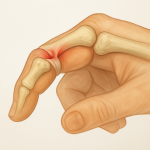
Xanthomas are localized collections of lipid-rich foam cells, usually found within the skin, subcutaneous tissue, or tendons. Among these, tendon xanthomas are a unique subset that occur within tendinous structures, most commonly in the Achilles tendon and the extensor tendons of the hands. These lesions are strongly associated with disorders of lipid metabolism, especially familial hypercholesterolemia (1). Though benign in nature, tendon xanthomas are often a visible marker of an underlying systemic condition that significantly elevates the risk of cardiovascular disease.
Tendon xanthomas are not just a cosmetic concern but are clinical indicators that warrant thorough investigation into lipid profiles and potential systemic risk. Their presence can serve as a critical diagnostic clue in detecting familial or secondary hyperlipidemias, especially in patients with no overt cardiovascular symptoms (2).
Symptoms
Tendon xanthomas typically present as (1).
- Firm, painless nodules or swellings along tendons
- Most commonly located at the Achilles tendon, patellar tendon, and extensor tendons of the hands
- Gradually increasing in size over time
- Non-tender and mobile with the tendon structure
- Occasionally associated with limited range of motion or discomfort with activity, especially when large
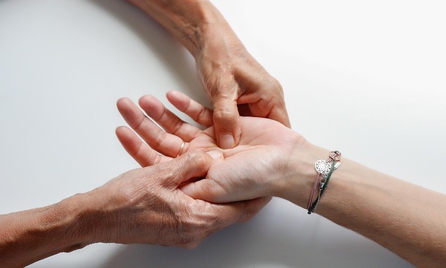
In rare cases, individuals may report mechanical issues such as difficulty walking (when in the Achilles tendon) or discomfort with gripping (when in hand tendons) (3).
Causes
The formation of tendon xanthomas is driven by the accumulation of lipids within macrophages, which then transform into foam cells and cluster in connective tissues, particularly tendons (2). This occurs primarily due to
- Elevated levels of low-density lipoprotein (LDL) cholesterol in the bloodstream
- Defects in the metabolism or clearance of lipids
- Genetic disorders, especially
- Familial hypercholesterolemia (FH)
- Type III hyperlipoproteinemia (familial dysbetalipoproteinemia)
These lipoprotein abnormalities lead to cholesterol being deposited in sites of slow turnover, such as tendons (1).
Risk Factors
Several risk factors predispose individuals to tendon xanthomas (2):
- Genetic Lipid Disorders
- Familial hypercholesterolemia is the most significant risk factor
- Homozygous FH leads to earlier and more extensive xanthoma development compared to heterozygous forms
- High LDL Cholesterol
- Uncontrolled LDL levels greatly enhance the likelihood of cholesterol deposition in tendons
- Age
- Tendon xanthomas are more prevalent in adults but can appear earlier in severe familial disorders
- Lack of Treatment or Poor Compliance
- Untreated or poorly managed hyperlipidemia significantly raises the risk of xanthoma formation
- Family History
- A history of hypercholesterolemia or xanthomas in close relatives is often present in affected individuals
- Sedentary Lifestyle and Poor Diet
- While not direct causes, they can aggravate existing lipid disorders, indirectly contributing to xanthoma development
Diagnosis
The diagnosis of tendon xanthomas involves a combination of clinical examination, imaging, and laboratory evaluation (3):
- Clinical Assessment
- Palpation of firm, non-tender, slow-growing masses along tendons
- Family history of lipid disorders or early cardiovascular events is often present
- Lipid Profile
- Elevated total cholesterol and LDL levels
- May also reveal abnormalities in triglycerides or HDL depending on the type of lipid disorder
- Genetic Testing
- Useful for confirming familial hypercholesterolemia
- Imaging Studies
- Ultrasound: Shows hypoechoic, thickened tendon with internal echoes
- MRI: Offers detailed imaging showing infiltrative lesions with altered tendon signal
- X-rays: May reveal soft tissue masses, although less specific
- Histopathological Examination
- Rarely needed unless the lesion is excised; confirms the presence of foam cells and lipid deposits
Treatment Options
Treatment focuses primarily on managing the underlying lipid disorder and monitoring or removing the xanthomas based on clinical necessity (2).
- Medical Management
- Lipid-Lowering Agents
- Statins (e.g., atorvastatin, rosuvastatin) to reduce LDL cholesterol
- Ezetimibe to inhibit cholesterol absorption
- PCSK9 inhibitors for resistant cases or familial hypercholesterolemia
- Fibrates or niacin if triglycerides are elevated
- Lifestyle Modifications
- Low-fat, high-fiber diet
- Regular aerobic exercise
- Smoking cessation
- Weight management
- Surgical Management
- Indicated when xanthomas cause:
- Significant cosmetic concern
- Functional impairment
- Pain or pressure symptoms
- Excision can be performed, though recurrence is possible without lipid control
- Experimental Therapies
- Newer agents like bempedoic acid or gene therapy are under evaluation for lipid disorders and may benefit in long-term control (1).
Living With or Prevention
Living with tendon xanthomas requires consistent and long-term management of lipid levels to prevent progression and associated cardiovascular complications (3).
Living With Tendon Xanthomas
- Regular monitoring of lipid profile and cardiovascular health
- Physical therapy if large xanthomas impair mobility
- Psychological support may be helpful due to cosmetic concerns or chronic disease burden
- Awareness of signs of cardiovascular events (e.g., chest pain, shortness of breath)
Prevention
- Early screening in individuals with family history of hyperlipidemia
- Genetic counseling for families with familial hypercholesterolemia
- Aggressive lipid control in high-risk individuals
- Encourage early lifestyle changes and adherence to medication regimens
References
- Oliva F, Marsilio E, Mastrodonato F, Migliorini F, Maffulli N. Minimally invasive excision and reconstruction of Achilles tendon xanthoma using free autologous semitendinosus tendon transfer: a surgical technique. J Orthop Surg Res. 2023 Apr 4;18(1):274. doi: 10.1186/s13018-023-03757-x. PMID: 37013640; PMCID: PMC10071761.
- Köroğlu M, Karakaplan M, Gündüz E, Kesriklioğlu B, Ergen E, Aslantürk O, Özdemir ZM. Cerebrotendinous Xanthomatosis patients with late diagnosed in single orthopedic clinic: two novel variants in the CYP27A1 gene. Orphanet J Rare Dis. 2024 Feb 9;19(1):53. doi: 10.1186/s13023-024-03082-4. PMID: 38336741; PMCID: PMC10858589.
- Zahradnik TM, Cresswell M, Squier K, Waugh C, Brunham L, Screen H, Scott A. Can Achilles tendon xanthoma be distinguished from Achilles tendinopathy using Dixon method MRI? A cross-sectional exploratory study. BMC Musculoskelet Disord. 2021 Jul 16;22(1):627. doi: 10.1186/s12891-021-04494-0. PMID: 34271888; PMCID: PMC8285885.