The glenoid is the shallow, cup-like socket of the scapula (shoulder blade) that holds the head of the humerus (upper arm bone), forming the ball-and-socket joint of the shoulder. A glenoid fracture refers to a break in this socket, which is an uncommon but serious type of shoulder injury. Although shoulder fractures are relatively common, glenoid fractures represent only a small percentage of them (1). Because the glenoid is crucial for maintaining shoulder stability and mobility, fractures in this area can severely affect arm function, range of motion, and quality of life if not diagnosed and managed properly. These injuries often occur in association with high-energy trauma, such as motor vehicle accidents, sports injuries, or falls (2).
Symptoms
The symptoms of a glenoid fracture can vary depending on the severity of the injury and whether the fracture is displaced (bones have shifted out of place) or non-displaced (2). Common symptoms include
- Severe pain in the shoulder, especially when moving the arm.
- Swelling and bruising around the shoulder joint.
- Restricted range of motion, making it difficult or impossible to lift the arm.
- Visible deformity in cases of displaced fractures.
- Grinding or clicking sensation during shoulder movement.
- Instability or slipping feeling in the joint if the fracture affects the socket’s shape.
- Numbness or tingling in the arm if nerves around the shoulder are compressed.
Because these symptoms can resemble other shoulder injuries like dislocations or rotator cuff tears, proper medical evaluation is essential.
Causes
Glenoid fractures typically result from direct or high-impact trauma (1). The most common causes include:
- Motor vehicle accidents: Sudden impact can drive the head of the humerus into the glenoid cavity, causing a break.
- Sports injuries: Contact sports such as football, rugby, and wrestling increase the risk of shoulder trauma.
- Falls: Landing directly on the shoulder or with an outstretched arm can transmit force to the socket, leading to a fracture.
- Direct blows: Accidents such as workplace injuries or heavy object impacts may directly fracture the glenoid.
- Seizures or electrical shocks: In rare cases, intense muscle contractions during seizures can exert enough force to fracture the glenoid (2).
Risk Factors
While anyone can suffer a glenoid fracture, certain factors increase the likelihood
- Participation in contact or high-impact sports: Athletes are more exposed to traumatic injuries.
- Occupational hazards: Jobs involving heavy machinery or risk of falling raise susceptibility (3).
- Age: Older adults with weakened bone density (osteoporosis) are more prone to fractures even with lower-impact falls.
- History of shoulder injuries: Previous dislocations or fractures may weaken the glenoid structure.
- High-risk behaviors: Not wearing seatbelts, extreme sports, or unsafe work practices can elevate risk.
Diagnosis
Accurate diagnosis is critical to ensure effective treatment and prevent long-term complications such as chronic instability or arthritis (1). Doctors generally use the following methods
- Physical examination: Assessing pain levels, range of motion, deformity, and shoulder stability.
- X-rays: The initial imaging test to identify the presence and type of fracture.
- CT scans (Computed Tomography): Provide detailed cross-sectional images to evaluate the exact location, shape, and displacement of the fracture.
- MRI scans (Magnetic Resonance Imaging): Useful in assessing damage to surrounding soft tissues like ligaments, cartilage, and rotator cuff muscles (2).
- Arthroscopy: In rare cases, a minimally invasive surgical camera may be used to assess the joint directly (1).
Glenoid fractures are classified based on their location and pattern (e.g., rim fractures, fossa fractures, or fractures extending into the scapular body), which guides treatment decisions.
Treatment Options
Treatment for a glenoid fracture depends on the type, size, and displacement of the fracture, as well as the patient’s overall health and activity level (3).
Non-Surgical Treatment
For non-displaced or minimally displaced fractures:
- Immobilization: A sling or shoulder immobilizer keeps the arm stable for several weeks.
- Pain management: Medications and cold compresses help reduce pain and swelling (1).
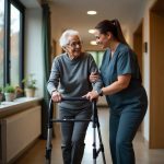
- Physical therapy: Once healing begins, guided exercises restore strength, mobility, and stability.
Surgical Treatment
Surgery is necessary when the fracture is displaced, involves a large portion of the glenoid surface, or causes instability. Common procedures include
- Open Reduction and Internal Fixation (ORIF): The fractured pieces are realigned and fixed with plates, screws, or pins.
- Arthroscopic surgery: Minimally invasive techniques may be used for small rim fractures (1).
- Bone grafting: In cases with bone loss, grafts may be used to restore joint stability.
- Shoulder replacement: In rare, severe fractures with extensive damage, partial or total shoulder replacement may be considered (2).
Post-surgical rehabilitation is crucial for regaining full function. This typically includes physical therapy, gradual strengthening exercises, and regular follow-ups with the surgeon.
Living With or Prevention
Recovery from a glenoid fracture can take several months, and lifestyle adjustments may be necessary during this period (2).
Living With a Glenoid Fracture
- Rehabilitation commitment: Consistently following prescribed physical therapy is essential for preventing stiffness and restoring mobility.
- Activity modification: Patients may need to avoid heavy lifting, contact sports, or high-impact activities until full healing occurs.
- Pain management: Use of prescribed medications, heat therapy, or ice packs can help with residual pain.
- Monitoring for complications: Watch for signs of arthritis, chronic pain, or instability, which may develop later.
Prevention
While not all glenoid fractures can be prevented, certain measures can reduce the risk
- Use protective gear: Helmets, shoulder pads, and safety harnesses in sports and occupations can lower injury chances (3).
- Fall prevention: Older adults should take steps to reduce fall risk through balance exercises, adequate lighting, and home safety measures.
- Strength training: Building shoulder and upper body strength enhances joint stability.
- Safe driving habits: Wearing seatbelts and avoiding reckless driving reduces risk in vehicle accidents.
- Bone health maintenance: Adequate calcium, vitamin D intake, and lifestyle habits that promote bone density help reduce fracture susceptibility.
References
- Chalidis B, Papadopoulos PP, Papadopoulos P, Pitsilos C. The Role of Arthroscopy in Contemporary Glenoid Fossa Fracture Fixation. Diagnostics (Basel). 2024 Apr 26;14(9):908. doi: 10.3390/diagnostics14090908. PMID: 38732322; PMCID: PMC11083719.
- Zhang X, Jiang Z, Chu FL, Jia DL, Han XG, Li XY, Zhao YF, Wang HB, Wu B. Comparative Study of Trans-Axillary Approach and Delto-Pectoral Approach to the Treatment of Ideberg Types I and II Scapular Glenoid Fractures. Orthop Surg. 2025 May;17(5):1349-1358. doi: 10.1111/os.70012. Epub 2025 Mar 2. PMID: 40024270; PMCID: PMC12050170.
- Pires RE, Giordano V, de Souza FSM, Labronici PJ. Current challenges and controversies in the management of scapular fractures: a review. Patient Saf Surg. 2021 Jan 6;15(1):6. doi: 10.1186/s13037-020-00281-3. PMID: 33407725; PMCID: PMC7789406.