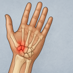
Synovial chondromatosis is a rare, benign (non-cancerous) joint disorder characterized by the formation of multiple cartilaginous nodules in the synovium—the thin membrane lining the joints. Over time, these nodules can detach and become loose bodies within the joint space, leading to pain, swelling, and decreased mobility. The condition most commonly affects large joints, especially the knee, although it can also involve the hip, elbow, shoulder, or ankle (1).
This disease primarily affects adults between the ages of 30 and 50 and tends to be more common in men than women. While synovial chondromatosis is not considered a life-threatening condition, it can significantly impair quality of life due to joint discomfort and functional limitations (1).
Symptoms
The clinical presentation of synovial chondromatosis varies depending on the joint involved and the extent of the disease. Common symptoms include (1):
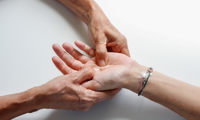
- Joint Pain: Persistent, aching pain that worsens with activity.
- Swelling: Visible or palpable swelling around the affected joint.
- Reduced Range of Motion: Stiffness and difficulty moving the joint fully.
- Locking or Catching Sensation: Caused by loose bodies interfering with joint movement (2).
- Joint Instability: In some cases, the joint may feel weak or unstable.
- Crepitus: A grinding or popping sound may be heard or felt during joint movement.
Symptoms typically develop gradually and may mimic those of other joint conditions, such as arthritis, making early diagnosis challenging.
Causes
The exact cause of synovial chondromatosis is unknown. It is generally considered to be idiopathic, meaning it arises spontaneously without a known reason. However, the condition involves metaplasia, a process in which synovial cells transform into cartilage-producing cells (2). This abnormal cellular behavior leads to the development of cartilaginous nodules within the joint lining.
There are two recognized forms of synovial chondromatosis (1):
- Primary Synovial Chondromatosis: Occurs independently and without any underlying joint disease. It is typically a monoarticular condition (affecting one joint).
- Secondary Synovial Chondromatosis: Develops in response to pre-existing joint abnormalities such as osteoarthritis, trauma, or inflammation. In this form, the cartilaginous nodules result from chronic joint irritation or damage.
Risk Factors
Several factors may increase the likelihood of developing synovial chondromatosis, including (3):
- Age: Most common in individuals aged 30 to 50.
- Gender: Men are affected more frequently than women.
- Joint Trauma: Previous injury to the joint may trigger abnormal synovial cell behavior.
- Chronic Joint Disorders: Conditions like osteoarthritis can predispose individuals to the secondary form.
- Repetitive Joint Use: Occupations or sports involving repeated joint stress may increase risk.
Although the condition is rare, awareness of these risk factors can prompt earlier investigation and diagnosis in symptomatic individuals.
Diagnosis
Diagnosing synovial chondromatosis involves a combination of clinical evaluation, imaging studies, and sometimes histological confirmation. The diagnostic process includes (2):
- Medical History and Physical Examination: The doctor will assess symptoms, duration, and impact on function, along with a physical exam to evaluate joint swelling, tenderness, and mobility.
- Imaging Techniques:
- X-rays: May show multiple calcified nodules within the joint in advanced stages.
- MRI (Magnetic Resonance Imaging): More sensitive for early disease detection and assessing non-calcified nodules.
- CT Scans: Provide detailed views of joint structures and may be used to assess the extent of calcification.
- Joint Aspiration: In some cases, joint fluid may be sampled to rule out infection or other inflammatory conditions.
- Biopsy: If malignancy is suspected or diagnosis is uncertain, surgical removal and histopathological examination of a nodule may be necessary.
Treatment Options
Treatment for synovial chondromatosis is largely surgical, aimed at relieving symptoms and preventing joint damage. The approach depends on disease severity, joint involved, and patient factors (3).
- Non-Surgical Management:
- Reserved for mild or asymptomatic cases.
- Includes NSAIDs (non-steroidal anti-inflammatory drugs) to manage pain and inflammation.
- Physical therapy to maintain joint function and strength.
- Surgical Treatment:
- Arthroscopic Removal: A minimally invasive procedure to remove loose bodies and sometimes perform a partial synovectomy (removal of affected synovial lining).
- Open Surgery: Required in cases with extensive disease, large nodules, or recurrent episodes. Complete synovectomy may be performed to reduce recurrence risk.
- Joint Replacement (Arthroplasty): Considered in advanced cases with significant joint damage or arthritis.
Surgical outcomes are generally favorable, though recurrence can occur, especially if all affected synovial tissue is not removed.
Living With or Prevention
Living With Synovial Chondromatosis
- While not preventable, individuals can take steps to manage symptoms and maintain joint health.
- Regular Exercise: Low-impact activities like swimming or cycling help maintain joint mobility.
- Weight Management: Reduces stress on weight-bearing joints.
- Pain Management: Use of prescribed medications and supportive therapies (e.g., hot/cold packs).
- Post-Surgical Rehabilitation: Crucial for recovery; includes physiotherapy to restore strength and function.
Monitoring
- Regular follow-ups are important, especially post-surgery, to detect recurrence or complications (3).
- MRI may be repeated periodically in cases of primary synovial chondromatosis due to the risk of recurrence.
Prevention
- There are no established prevention strategies for primary synovial chondromatosis.
- Managing underlying joint conditions and avoiding repetitive joint trauma may help reduce the risk of secondary synovial chondromatosis.
References
- Monestier L, Riva G, Stissi P, Latiff M, Surace MF. Synovial chondromatosis of the foot: Two case reports and literature review. World J Orthop. 2019 Nov 18;10(11):404-415. doi: 10.5312/wjo.v10.i11.404. PMID: 31840021; PMCID: PMC6908443.
- Sviland L, Malcolm AJ. Synovial chondromatosis presenting as painless soft tissue mass–a report of 19 cases. Histopathology. 1995 Sep;27(3):275-9. doi: 10.1111/j.1365-2559.1995.tb00221.x. PMID: 8522293.
- Lasmar NP, Vieira RB, Rosa Jde O, Lasmar RC, Scarpa AC. SYNOVIAL CHONDROMATOSIS. Rev Bras Ortop. 2015 Dec 12;45(5):490-2. doi: 10.1016/S2255-4971(15)30441-9. PMID: 27047814; PMCID: PMC4799217.