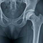
A Perthes lesion is a specific type of shoulder injury involving the glenohumeral (shoulder) joint. It is characterized by the avulsion or tearing of the anteroinferior glenohumeral ligament (AIGHL) from the glenoid (the socket of the shoulder joint), while the periosteum (the fibrous layer covering bone) remains intact and stripped medially (1). This type of lesion is commonly associated with anterior shoulder dislocations and is often mistaken for other shoulder injuries such as Bankart lesions or ALPSA (Anterior Labroligamentous Periosteal Sleeve Avulsion) lesions. Early and accurate diagnosis of a Perthes lesion is crucial to avoid chronic instability and long-term joint dysfunction (1).
Symptoms
The clinical presentation of a Perthes lesion can be subtle, especially in the early stages. Symptoms often overlap with other forms of shoulder instability. Common signs and symptoms include:
- Recurrent shoulder dislocations or subluxations (partial dislocations)
- Persistent shoulder pain, especially during activities involving arm elevation or rotation
- Feeling of instability or “giving way”
- Weakness in the shoulder
- Decreased range of motion, particularly during external rotation
- Clicking, popping, or catching sensations in the shoulder joint
These symptoms may vary depending on the severity of the lesion and whether it is accompanied by other structural injuries.
Causes
The most common cause of a Perthes lesion is trauma, specifically anterior shoulder dislocation. When the shoulder is forced into extreme abduction and external rotation, the humeral head (ball of the shoulder joint) can dislocate forward, placing significant stress on the soft tissues that stabilize the joint (1).
During such a dislocation:
- The anteroinferior glenohumeral ligament may tear away from the glenoid rim.
- The periosteum, although not completely torn, is peeled away medially.
This unique injury pattern results in a lesion that can be difficult to detect through standard imaging techniques.
Risk Factors
Several risk factors may predispose an individual to developing a Perthes lesion:
- History of shoulder dislocation – Especially if the dislocation occurred at a young age or was left untreated.
- Repetitive overhead activity – Athletes such as swimmers, baseball pitchers, and volleyball players are more susceptible due to repeated stress on the shoulder joint.
- Contact sports – Football, rugby, and martial arts increase the risk due to the high likelihood of shoulder trauma.
- Joint hypermobility or laxity – Individuals with more flexible joints may be more prone to ligamentous injuries.
- Poor rehabilitation after initial shoulder dislocation – Inadequate healing can lead to residual instability and further injuries.
- Previous shoulder surgery or injury – Prior trauma or surgical intervention can compromise the integrity of the joint and its stabilizing structures.
Diagnosis
Diagnosing a Perthes lesion requires a high degree of clinical suspicion, particularly in patients with persistent shoulder instability after an initial traumatic event (1). Diagnosis involves:
Clinical Examination
- Positive apprehension test (patient feels the shoulder may dislocate when externally rotated and abducted).
- Load and shift test to assess joint stability.
Imaging
- MRI (Magnetic Resonance Imaging) is the most useful tool. It helps visualize soft tissue injuries, including the glenoid labrum and ligamentous structures. An MRI with intra-articular contrast (MR arthrogram) significantly improves detection.
- CT scan with 3D reconstruction may be used to assess associated bony injuries.
- Arthroscopy, a minimally invasive surgical procedure, may be necessary for definitive diagnosis and is often used to confirm ambiguous MRI findings.
Because a Perthes lesion can mimic other labral injuries, correct interpretation by an experienced radiologist or orthopedic surgeon is essential.
Treatment Options
The management of Perthes lesions depends on the severity of the lesion and the level of instability experienced by the patient (2).
Non-Surgical Treatment
- Rest and Immobilization: Initially, the shoulder may be immobilized using a sling to allow soft tissue healing.
- Physical Therapy: Focuses on strengthening the rotator cuff and scapular stabilizers to restore shoulder stability.
- Activity Modification: Avoidance of activities that provoke symptoms or risk re-injury.
However, non-surgical treatment is often not sufficient, particularly in active individuals or athletes (2).
Surgical Treatment
When conservative management fails or if the lesion is identified early in active patients, surgical intervention may be recommended. The main surgical approach is arthroscopic repair, which involves:
- Reattaching the avulsed ligament to the glenoid rim.
- Securing the labrum and reinforcing the shoulder capsule using suture anchors.
Arthroscopy offers the advantage of less postoperative pain, shorter hospital stays, and quicker return to activity. Post-surgical rehabilitation is critical and involves a structured program that gradually progresses from passive range of motion to active strengthening (2).
Living With or Prevention
Living with a Perthes lesion, especially if untreated or misdiagnosed, can lead to chronic shoulder instability, reduced athletic performance, and early-onset osteoarthritis. Therefore, timely and accurate treatment is crucial for long-term joint health (3).
Tips for Prevention and Long-term Care
- Proper Training Techniques: Athletes should use correct form and avoid overuse.
- Strength Training: Building strength in the rotator cuff and shoulder stabilizers can help prevent dislocations.
- Protective Gear: In contact sports, using shoulder pads can help reduce the impact of collisions.
- Gradual Return to Activity: After treatment, athletes should return to sports only after regaining full strength and stability.
- Regular Follow-ups: Monitoring with periodic physical assessments or imaging can help identify any recurrence of instability.
References
- Rodríguez-Olivas AO, Hernández-Zamora E, Reyes-Maldonado E. Legg-Calvé-Perthes disease overview. Orphanet J Rare Dis. 2022 Mar 15;17(1):125. doi: 10.1186/s13023-022-02275-z. PMID: 35292045; PMCID: PMC8922924.
- Anderson LA, Erickson JA, Severson EP, Peters CL. Sequelae of Perthes disease: treatment with surgical hip dislocation and relative femoral neck lengthening. J Pediatr Orthop. 2010 Dec;30(8):758-66. doi: 10.1097/BPO.0b013e3181fcbaaf. PMID: 21102198; PMCID: PMC3031125.
- Clohisy JC, Nepple JJ, Ross JR, Pashos G, Schoenecker PL. Does surgical hip dislocation and periacetabular osteotomy improve pain in patients with Perthes-like deformities and acetabular dysplasia? Clin Orthop Relat Res. 2015 Apr;473(4):1370-7. doi: 10.1007/s11999-014-4115-7. PMID: 25560960; PMCID: PMC4353550.